
Imaging cardiology, also known as cardiovascular imaging or cardiac imaging, is a specialized field within cardiology that focuses on using various imaging techniques to visualize and evaluate the structure, function, and diseases of the heart and blood vessels. Imaging cardiology in Vellore encompasses a range of non-invasive and invasive imaging modalities that aid in the diagnosis, treatment planning, and monitoring of cardiovascular conditions.
Imaging cardiologists are physicians who have specialized training and expertise in the interpretation and application of cardiovascular imaging techniques. They play a crucial role in providing accurate diagnoses, guiding treatment decisions, and monitoring the effectiveness of interventions.
Imaging cardiology employs several imaging modalities to obtain detailed images of the heart and blood vessels. These techniques allow imaging cardiologists to assess various aspects of cardiovascular health, including structure, function, blood flow, and tissue characteristics. Some commonly used modalities for imaging cardiology in Vellore include:
Role of Imaging Cardiology in Vellore
At our cardiovascular center, our dedicated team of cardiologists plays a pivotal role in safeguarding and enhancing your heart health. With a commitment to excellence, our cardiologists bring a wealth of expertise to the table, contributing to every aspect of cardiovascular care.
Echocardiography
Echocardiography is a non-invasive imaging technique that utilizes ultrasound waves to create real-time images of the heart. It provides valuable information about the heart’s structure, function, blood flow patterns, and valves. Echocardiography can diagnose conditions such as heart valve disorders, heart muscle abnormalities, congenital heart defects, and fluid accumulation around the heart (pericardial effusion).
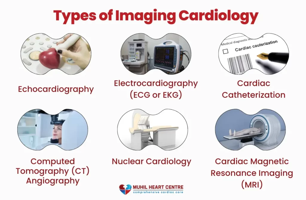
Cardiac Magnetic Resonance Imaging (MRI)
Cardiac MRI employs radio waves and a strong magnetic field to generate detailed images of the heart. It provides information about the heart’s structure, function, blood flow, and tissue characteristics. Cardiac MRI is particularly useful for evaluating conditions such as heart muscle damage (myocardial infarction), heart tumors, congenital heart defects, and assessing blood flow in the heart and blood vessels.
Computed Tomography (CT) Angiography
CT angiography involves the use of X-rays and computer technology to create three-dimensional images of the heart and blood vessels. It helps visualize the coronary arteries, assess the extent of coronary artery disease, and identify areas of arterial blockages or stenosis. CT angiography is valuable in pre-operative planning for coronary interventions and guiding treatment decisions.
Nuclear Cardiology
Nuclear cardiology utilizes small amounts of radioactive materials, known as radiotracers, to evaluate blood flow, heart function, and metabolism. Techniques such as single-photon emission computed tomography (SPECT) and positron emission tomography (PET) used for imaging cardiology in Vellore can help assess myocardial perfusion (blood flow to the heart muscle), viability, and detect the presence of conditions like cardiac amyloidosis or inflammation.
Cardiac Catheterization and Angiography
Cardiac catheterization is an invasive procedure that involves inserting a thin tube (catheter) into the heart and blood vessels. During the procedure, contrast dye is injected, and X-ray imaging (angiography) is performed to visualize the coronary arteries and assess blood flow. Cardiac catheterization provides important information about the presence and severity of blockages or narrowing in the arteries, blood pressure measurements within the heart chambers, and oxygen levels.
Stress Testing
Stress testing evaluates the heart’s response to physical exercise or medications. It is often combined with imaging techniques such as echocardiography, nuclear imaging, or cardiac MRI to assess the heart’s function and detect any abnormalities under stress conditions. Stress testing can help diagnose coronary artery disease, evaluate exercise tolerance, and determine the need for further interventions.
Intravascular Imaging
Intravascular imaging techniques, such as intravascular ultrasound (IVUS) and optical coherence tomography (OCT), are used during cardiac catheterization procedures. This technique of imaging cardiology in Vellore provides high-resolution images of the inside of the coronary arteries, helping assess plaque buildup, stenosis, and guide interventions such as stent placement.
Read more about Heart hole treatment in Vellore
Role of our Cardiologist
Diagnosis and Evaluation
Our imaging cardiologist is skilled in interpreting imaging studies to accurately diagnose and evaluate cardiovascular conditions. We analyze images obtained from different imaging modalities and integrate them with clinical data to make informed diagnostic decisions.
Through careful evaluation, we can identify abnormalities in the heart’s structure, function, blood flow, and tissue characteristics, leading to accurate diagnoses of conditions such as coronary artery disease, heart valve disorders, heart failure, arrhythmias, and congenital heart defects.
Treatment Planning and Guidance
Our imaging cardiologist provides critical information for treatment planning and guiding interventions. By assessing the extent and severity of cardiovascular disease, we assist in determining the most appropriate treatment strategies.
Interventional Procedure Guidance
During interventional procedures, such as cardiac catheterization or percutaneous coronary interventions, imaging cardiologists play a crucial role in guiding the procedures. We use real-time imaging cardiology in Vellore to visualize the coronary arteries, assess the placement of devices (such as stents), and ensure the accurate delivery of therapies. With our expertise in intravascular imaging, we can provide detailed guidance to interventional cardiologists, facilitating precise interventions.
Monitoring and Follow-Up
Imaging cardiologists are involved in monitoring patients’ progress and assessing the effectiveness of treatments. Through follow-up imaging studies, we can evaluate changes in the heart’s structure, function, and blood flow over time. By comparing current imaging results with previous ones, we can determine whether treatments are successful, detect disease progression, or identify potential complications.

Multidisciplinary Collaboration
Imaging cardiologists work closely with other healthcare professionals, including cardiologists, interventional cardiologists, cardiac surgeons, radiologists, and referring physicians. We collaborate as part of a multidisciplinary team to ensure comprehensive and patient-centered care. By sharing expertise and insights, we collectively determine the most appropriate diagnostic and treatment pathways for patients.
Patient Education and Communication
The expert in imaging cardiology in Vellore plays an essential role in patient education and communication. We explain imaging findings, treatment options, and the significance of diagnostic tests to patients and their families. We help patients understand their cardiovascular conditions, potential risks, and the importance of adhering to recommended treatments. By providing clear explanations and addressing patient concerns, we empower individuals to actively participate in their own healthcare journey.
FAQ
What types of cardiac imaging services are available in the Cardiology Imaging department in Vellore?
The Cardiology Imaging department in Vellore offers a wide range of advanced imaging techniques to assess heart health. These may include echocardiography, cardiac MRI (Magnetic Resonance Imaging), CT (Computed Tomography) angiography, and nuclear cardiology. Each of these imaging modalities serves different purposes in diagnosing and monitoring heart conditions.
How can I prepare my child or myself for a cardiac imaging procedure in Vellore?
Preparation for cardiac imaging procedures can vary depending on the specific test being performed. Patients are typically advised to follow fasting instructions, avoid certain medications, and wear comfortable clothing. It’s important to consult with the healthcare provider or imaging center well in advance to receive detailed instructions tailored to your specific procedure.
Are the imaging facilities in Vellore equipped with the latest technology for accurate cardiac diagnosis?
Yes, the cardiac imaging facilities in Vellore are equipped with state-of-the-art technology to ensure accurate and precise cardiac diagnoses. These facilities often invest in cutting-edge equipment and employ experienced radiologists and technologists who specialize in cardiovascular imaging. Patients can expect high-quality, advanced imaging services for the most accurate assessment of their heart health.
Conclusion
To conclude, imaging cardiology in Vellore plays a crucial role in diagnosing and managing cardiovascular diseases, providing valuable insights for accurate diagnosis, treatment planning, and improved patient outcomes. It continues to advance healthcare by enhancing our understanding of the heart’s structure and function.
Read also Preventive Cardiology in Vellore.